 JawAtlas
Reference
CBCT glossary for dental students
Every term you are likely to meet while learning dental CBCT, and every term JawAtlas uses, defined in one to three sentences and grouped so you can read one section at a time.
Terms are grouped by where you meet them: in the scanner, in the geometry of the slices, on the screen, in the reconstructions, in the files, in the anatomy, and in the app. For the longer story behind any group, follow the links at the end of each section.
Acquisition
- Cone beam
- The X-ray geometry used by CBCT: a cone or pyramid of X-rays that covers the whole region in one exposure and lands on a flat panel detector. Medical CT uses a thin fan instead and builds the volume slice by slice.
- Voxel
- One small box of the three dimensional volume, holding one grey value. It is the 3D equivalent of a pixel, and everything you see on a slice is a sheet of voxels.
- Isotropic
- Voxels that have the same size in every direction. Most dental CBCT volumes are isotropic or nearly so, which is why a coronal or sagittal slice looks as sharp as an axial one.
- Field of view (FOV)
- The size of the cylinder or sphere the scanner captures, usually given as diameter by height in millimetres. A small FOV covers a few teeth; a large one covers both jaws. The demo scan is about 127 by 80 mm, centred on the mandible.
- Reconstruction
- The computation that turns the few hundred projection images taken during the scan into a block of voxels. Several artifacts are born in this step.
- Beam hardening
- The X-ray beam loses its low energy photons as it passes through dense material, so it comes out harder than the reconstruction expects. The result is dark bands and bright streaks radiating from metal and dense enamel.
- Partial volume effect
- A voxel that contains two materials holds the average of both. Thin structures such as the lamina dura or a cortical plate look fainter than they are, or vanish, when they are thinner than the voxel.
- Motion artifact
- Blur and doubled outlines caused by the patient moving during the scan. It affects the whole volume, which separates it from local artifacts.
- Noise
- Random grain in the grey values, worse with low exposure and small voxels. It is uniform and has no direction, unlike streaks.
- Ring artifact
- Faint concentric circles on axial slices caused by a detector element that reads differently from its neighbours. They are centred on the axis of rotation and ignore anatomy.
- Scatter
- Photons deflected inside the patient that reach the detector in the wrong place. The flat panel in a CBCT unit catches a lot of scatter, which lowers contrast and makes grey values drift across the volume.
Read more: CBCT artifacts and field of view and voxel size.
Geometry and planes
- Axial
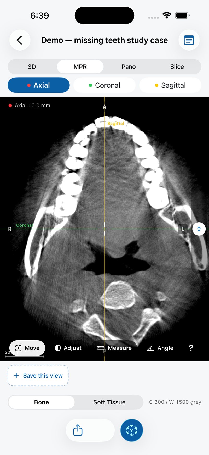
- A horizontal slice, as if looking at the jaw from above or below. By radiological convention it is viewed from the feet, so the patient's left is on the viewer's right. Red in JawAtlas.
- Coronal
- A vertical slice running from ear to ear, as if facing the patient. The patient's left is again on the viewer's right. Green in JawAtlas.
- Sagittal
- A vertical slice running from front to back, showing the patient in profile. Yellow in JawAtlas.
- Oblique
- Any slice that is not aligned with the three standard planes. The Slice stage in JawAtlas is a free oblique slice you can tilt in any direction.
- MPR (multiplanar reconstruction)
- Cutting the volume along planes other than the acquired one, usually the three standard planes shown side by side and linked by a shared point.
- Crosshair
- The marker of the single point in space that all MPR planes share. Move it in one plane and the other planes jump to the slices through that point. White in JawAtlas.
- Scout line
- A line drawn on one plane showing where another plane cuts through it, in the colour of the plane it represents. On the panoramic view, the yellow scout line sets where the cross-section is taken.
- Slab
- A slice with thickness: several neighbouring voxel layers combined into one image, by averaging or by taking the maximum. A thin slab shows detail; a thick slab shows more structure at once but blurs it.
- Radiological convention
- The agreement that axial and coronal images are displayed as if you were facing the patient, so the patient's left is on the right of the screen. Check the edge labels rather than assuming.
- Orientation labels
- The letters at the edges of a slice: R and L for the patient's right and left, A and P for anterior and posterior, H and F for head and feet. JawAtlas derives them from the orientation stored in the scan.
Read more: axial, coronal and sagittal planes, orientation conventions and tri-planar MPR.
Display
- Grey value
- The number stored in each voxel of a CBCT scan, which the display turns into a shade of grey. CBCT grey values are relative, not calibrated, so the same tissue can read differently on two scanners or in two parts of one scan.
- Hounsfield unit (HU)
- The calibrated density scale of medical CT, with water at 0 and air at minus 1000. CBCT does not produce true HU, which is why JawAtlas shows grey values and never HU.
- Window
- The range of grey values mapped from black to white. Everything below the range is black, everything above is white. Also called window width.
- Level
- The grey value at the centre of the window, also called the window centre. Raising the level makes the image darker overall; lowering it makes it brighter. JawAtlas shows both as "C" and "W" in grey values.
- Bone window
- A wide window centred on high grey values, so that bone and teeth show detail and soft tissue is uniformly dark. The Bone preset in JawAtlas.
- Soft tissue window
- A narrow window centred on soft tissue grey values, so that muscle, mucosa and air separate and bone is uniformly white. The Soft Tissue preset in JawAtlas.
- Adjust mode
- The JawAtlas toolbar mode in which a one finger drag changes the window: up and down for level, left and right for width.
Read more: window, level and grey values and adjusting window and level.
Reconstructions
- MIP (maximum intensity projection)
- A projection through the volume in which each pixel shows the brightest voxel along its line of sight. The result looks like a radiograph, with bone and teeth standing out and depth information lost.
- Volume rendering
- Drawing the whole block of voxels as a 3D object by giving each grey value a colour and an opacity and accumulating them along each line of sight. The 3D stage in JawAtlas.
- Transfer function
- The rule that maps grey values to colour and opacity in volume rendering. The Bone, Teeth and Soft tissue presets in JawAtlas are three transfer functions.
- Surface shading
- Lighting applied to a volume rendering so that surfaces facing the light look brighter, which gives the 3D view depth. The Shaded toggle in JawAtlas turns it on and off.
- Ray marching
- The method used to render a volume: for every pixel, a ray is stepped through the block of voxels, sampling and accumulating colour and opacity as it goes. JawAtlas does this on the GPU with Metal.
- Panoramic reconstruction
- A flat image built from CBCT by sampling the volume along a curve that follows the dental arch and averaging a slab of voxels around it. It resembles a panoramic radiograph but is computed from the scan, not acquired separately.
- Arch curve
- The curve you draw on an axial slice to tell the software where the dental arch runs. In JawAtlas you trace it with at least four taps in Edit arch, and it is stored with the case.
- Cross-section
- A slice taken at right angles to the arch curve at a chosen point along it. It shows the buccolingual dimension that the panoramic image cannot, and in JawAtlas it follows the yellow scout line on the pano.
Read more: panoramic reconstruction from CBCT and maximum intensity projection.
Files and data
- DICOM
- The standard file format and data model for medical images. A dental CBCT scan is usually exported as a folder of DICOM files, one per slice, each carrying the pixel data plus tags that describe it.
- Series
- The set of DICOM files that together make one volume. JawAtlas builds a series by sorting the slices physically along the slice direction and checking for gaps and duplicates; a series needs at least four slices.
- Transfer syntax
- The DICOM tag that says how the pixel data is encoded: uncompressed, deflated, RLE, lossless JPEG and so on. JawAtlas reads the uncompressed and lossless kinds and rejects lossy JPEG, JPEG 2000 and JPEG-LS files with a message.
- Pixel spacing
- The physical size of one pixel within a slice, in millimetres, stored in the DICOM header. Together with slice spacing it gives the voxel size and makes measurements possible.
- Slice thickness
- The thickness of tissue each slice represents, as stored in the header. In CBCT it usually equals the voxel size.
- Slice spacing
- The distance between the centres of neighbouring slices. JawAtlas measures it from the slice positions rather than trusting a single tag, which is how it detects gaps.
- ImageOrientationPatient
- The DICOM tag holding the direction cosines of the slice rows and columns relative to the patient. JawAtlas uses it to derive the R, L, A, P, H and F edge labels, so they stay correct on rotated or oblique volumes.
- ImagePositionPatient
- The DICOM tag holding the 3D position of the first voxel of each slice. Sorting slices by this position, along the slice normal, is how a volume is assembled in the right order.
- Rescale slope and intercept
- Two DICOM tags that convert stored pixel values to the scanner's output scale. JawAtlas handles files where they are missing.
- Multi-frame file
- A single DICOM file that holds every slice of the volume instead of one file per slice. JawAtlas imports both layouts.
- Anonymisation
- Removing identifying information such as name, ID and birth date from a scan. JawAtlas never stores these: the importer reads only a whitelist of technical tags, so identifiers never reach the app's storage.
- Case package
- A shared JawAtlas case, with its moments and notes, delivered through the share sheet or AirDrop. A folder of them can be batch-imported.
Read more: what is DICOM and importing DICOM from Files.
Anatomy landmarks
- Cortical bone
- The dense outer shell of bone, which appears bright white on CBCT. The outer surface of the mandible, the lower border and the walls of the sinus are cortical.
- Trabecular bone
- The spongy inner bone, a mesh of thin bright struts with dark marrow spaces between. Its texture is where many fine structures hide, and where partial volume does the most damage.
- Lamina dura
- The thin layer of dense bone lining the tooth socket, seen as a bright line around the root just outside the periodontal ligament space.
- Periodontal ligament space
- The thin dark line between the root surface and the lamina dura, where the ligament fibres sit. It is near the limit of what CBCT can resolve and is often partly lost to partial volume.
- Inferior alveolar canal
- The tunnel through the mandible that carries the inferior alveolar nerve and vessels, from the mandibular foramen to the mental foramen. A dark channel, often with a thin bright rim, running below the molar roots.
- Mandibular foramen
- The opening on the inner surface of the ramus where the inferior alveolar canal begins, roughly at the level of the occlusal plane, guarded by a small spur of bone called the lingula.
- Mental foramen
- The opening on the outer surface of the mandible, usually near the premolar apices, where the canal ends. On an axial slice it is a gap in the buccal cortex.
- Incisive canal
- In the maxilla, the canal behind the central incisors that opens at the incisive foramen in the palate and shows as a dark channel in the midline. The mandible has a smaller incisive canal too, the faint continuation of the inferior alveolar canal beyond the mental foramen.
- Maxillary sinus
- The large air space in the body of the maxilla above the premolar and molar roots, outlined by thin cortical walls. Air makes it almost black on every window.
- Nasal floor
- The bony floor of the nasal cavity, formed by the hard palate, seen on coronal slices as a bright line beside and usually above the sinus floor.
- Condyle
- The rounded head at the top of the posterior process of the ramus, which sits in the glenoid fossa of the temporal bone to form the temporomandibular joint.
- Coronoid process
- The thin, triangular process at the top of the anterior border of the ramus, in front of the condyle, where the temporalis muscle attaches.
- Mylohyoid ridge
- An oblique ridge on the inner surface of the mandibular body running down and forward from the molar region, seen on cross-sections as a bright bump on the lingual cortex.
Read more: finding the mandibular canal, the maxillary sinus and impacted third molars.
In JawAtlas
- Case
- One imported scan together with its arch curve, moments and notes. Cases live in the library and can be shared as a package or exported as a PDF summary.
- Moment
- A saved view of a case: the slice, plane, window and any mark you drew, with a caption. Moments replay in order like slides and travel with the case when you share it.
- Stage
- One of the four ways of looking at a case, chosen with the segmented control at the top: 3D, MPR, Pano and Slice.
- Slice view
- The stage that shows a single free oblique slice you can tilt in any direction, with orientation chips and presets such as Top.
- AR slice-through
- Anchoring the scan life-size on a flat surface and moving the device through it, so the cross-section follows the device in real time. It needs a physical iPhone or iPad with ARKit.
- Demo scan
- The CBCT case bundled with the app and seeded on first launch: case M29 from the openly licensed dataset by Feng and colleagues (CC BY 4.0), a mandible-centred volume with a full arch, both rami, a left posterior edentulous site and a traceable inferior alveolar canal. It can be restored from Settings.
- Measure and Angle
- Toolbar modes that read a length in millimetres between two taps, or an angle in degrees, in any of the three MPR panes. Both are calibrated from the DICOM spacing.
- Scale ruler
- The scale bar at the bottom left of the MPR pane and the pano cross-section (20 mm at a typical zoom; it picks 5, 10, 20, 50 or 100 mm to fit), there so you can judge size at a glance without measuring.
Read more: all features, about the demo scan and saving and replaying moments.
Questions students ask
What is the difference between a voxel and a pixel?
A pixel is one square element of a two dimensional picture. A voxel is one small box of a three dimensional volume. A CBCT scan is a block of voxels, and every slice you look at is a sheet of them.
Why does JawAtlas show grey values instead of Hounsfield units?
Hounsfield units are a calibrated scale that medical CT scanners produce, with water at 0 and air at minus 1000. CBCT grey values are not calibrated that way and drift with position, field of view and scatter, so showing them as HU would suggest an accuracy that is not there.
What is a moment in JawAtlas?
A saved view: the slice, plane, window and any mark you drew, with a caption. Moments can be replayed in order like slides, and they travel with the case when you share it.
What does MPR stand for?
Multiplanar reconstruction: cutting a three dimensional volume along planes other than the one it was acquired in, usually the axial, coronal and sagittal planes, which all share one crosshair point.
JawAtlas is an education and training aid. It is not a medical device and must not be used for diagnosis, treatment planning or any clinical decision.
Last reviewed 2026-10-13