 JawAtlas
Guide
Radiological orientation conventions on CBCT
After this guide you will be able to tell the patient's right from left in any CBCT view, explain why slices and panoramic images use different conventions, and avoid the side mistakes that catch most beginners.
Why axial slices are viewed from the feet
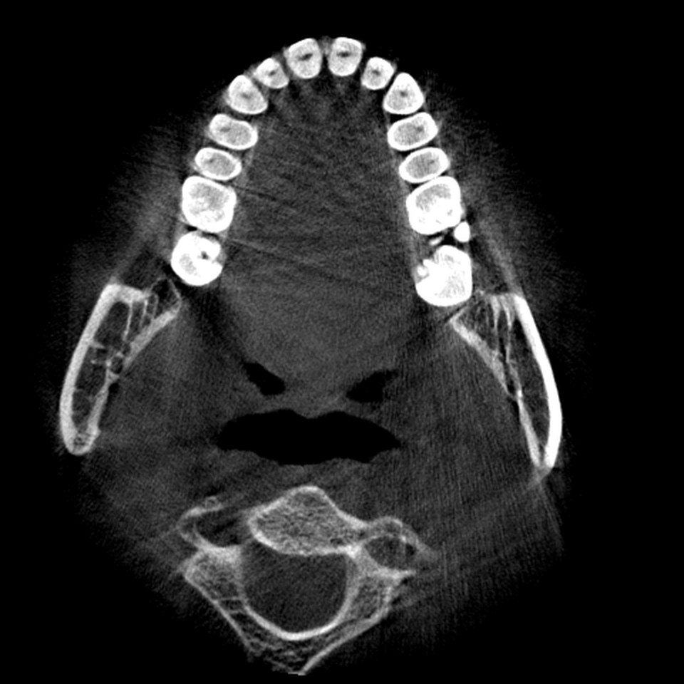
Radiology uses a single rule for cross-sectional images: an axial slice is displayed as if the viewer stood at the feet of a patient lying on their back and looked towards the head. From that position the front of the patient is at the top of the image and the patient's right side is on the viewer's left. The rule comes from medical CT, where the patient lies supine on the table, and it was kept by MRI and later by CBCT so that a reader moving between modalities never has to relearn sides.
Coronal slices follow the same idea. They are displayed as if the viewer faced the patient, so again the patient's right is on the viewer's left and the head is at the top. Sagittal slices are a profile view; in JawAtlas they are shown with anterior on the viewer's left and the head at the top. Dental CBCT scanners usually image a seated or standing patient rather than a supine one, but the display convention does not change with the scanner. It is a convention about the picture, not about the machine.
Patient left on the viewer's right
The easiest way to hold the convention in your head is to notice that it matches how you see a person standing in front of you. Face someone and their right hand is on your left. An axial or coronal slice is presented the same way, so the convention is not a mirror image, it is the normal view of a patient you are facing. What makes it feel wrong is that students often picture themselves looking down on the arch from above, as a dentist does at the chair. In that picture the patient's right is on your right. Radiology deliberately does not use that picture.
A second trap is the panoramic radiograph. It also follows the facing convention, so the patient's right is on the viewer's left, but because it shows the arch flattened from the front rather than cut from the feet, the teeth are arranged the other way up compared with an axial slice. A student who learned sides on panoramic films and then opens an axial stack finds the arch upside down and the sides apparently reversed. They are not reversed; the two views are related by a rotation, not a reflection.
The edge labels R, L, A, P, H and F
Because conventions are easy to forget and easy to break, every slice carries letters at its edges that state the direction the patient's body points at that edge. R and L are the patient's right and left. A and P are anterior and posterior. H and F are head and feet (some systems write S and I for superior and inferior instead). On an axial slice you should see A at the top, P at the bottom, R on the left and L on the right. On a coronal slice you should see H at the top, F at the bottom, R on the left and L on the right. On a sagittal slice in JawAtlas you see H at the top, F at the bottom, A on the left and P on the right.
The labels are the only reliable statement of orientation, so read them every time instead of trusting the shape of the image. JawAtlas draws them on all three MPR panes and on the free oblique Slice view, where they matter even more because the slice does not sit in a standard plane. The planes guide shows what each standard slice looks like with its labels.
The OPG convention for panoramic images
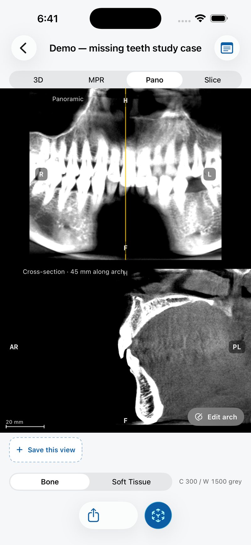
A conventional panoramic radiograph (OPG) is displayed as if you faced the patient: the patient's right side of the jaw is on the viewer's left, and the image reads like a portrait of the teeth. A panoramic reconstruction computed from a CBCT volume keeps the same convention so that it looks like the film students already know. In JawAtlas the Pano stage puts the patient's right on the viewer's left for this reason, even though the axial slice you traced the arch on had the patient's right on the left as well, just with anterior at the top.
When you move between the Pano stage and the MPR stage, the sides do not swap; what changes is the direction you are looking from. The yellow scout line on the pano and the cross-section it drives are both labelled, so you can always check. The CBCT versus panoramic radiograph guide covers what else differs between the two images.
How DICOM stores orientation
A viewer does not guess any of this. Each DICOM slice carries a tag called ImageOrientationPatient with six numbers: the first three are the direction cosines of the image rows and the last three are the direction cosines of the image columns, both expressed in the patient coordinate system, where x points to the patient's left, y points posterior and z points towards the head. A perfectly aligned axial slice has rows along x and columns along y, so its cosines are 1, 0, 0 and 0, 1, 0. If the scan was acquired tilted or the patient was turned, the cosines carry that tilt, and a viewer that reads them can still say with confidence which edge of the image is the patient's left. JawAtlas derives the R, L, A, P, H and F labels from these cosines, which is why they stay correct on oblique and rotated volumes. The DICOM guide explains the rest of the file.
Common student mistakes
The first mistake is assuming the chairside view. Students who picture the arch from above put the patient's right on their own right and then describe findings on the wrong side. The cure is to read the labels before you say a side out loud.
The second is carrying the panoramic picture into an axial stack and concluding that the arch is "upside down" or "mirrored". The arch is simply seen from a different direction. Trace the patient's right from the pano to the axial slice with the labels and the relationship becomes obvious after a few scans.
The third is trusting the image itself on a scan that was acquired tilted. A head that was rotated in the scanner can make the midline look off centre and one ramus look longer. The labels, computed from the DICOM cosines, stay correct even when the picture looks odd, and the free oblique Slice view can be tilted to put the midline back where you expect it. The reading routine makes orientation the first step for exactly these reasons.
Questions students ask
Why is the patient's left on the right of an axial CT or CBCT slice?
Axial slices are displayed as if you stand at the feet of a patient lying on their back and look towards the head. From that position the patient's right is on your left. The rule comes from medical CT and every cross-sectional modality keeps it so that sides never need relearning.
What do the letters R, L, A, P, H and F on a CBCT slice mean?
They mark the direction the patient's body points at that edge of the image: right, left, anterior, posterior, head and feet. Read them rather than assuming, because they are the only reliable statement of which side is which.
Why is a panoramic image the other way round from an axial slice?
A panoramic radiograph follows the OPG convention, which shows the patient as if you faced them from the front with their right on your left. An axial slice is viewed from the feet with anterior at the top, which puts the arch the other way up. Both agree that the patient's right is on the viewer's left when you face them.
What is ImageOrientationPatient in a DICOM file?
It is a tag holding six numbers: the direction cosines of the image rows and columns in the patient coordinate system. A viewer uses them to work out which way the patient's left, anterior and head point in each slice and to draw correct edge labels even when the volume was scanned tilted.
JawAtlas is an education and training aid. It is not a medical device and must not be used for diagnosis, treatment planning or any clinical decision.
Last reviewed 2026-10-13