 JawAtlas
Guide
Axial, coronal and sagittal planes on a dental CBCT
After this guide you will know what each of the three standard planes shows best on a jaw scan, how the slices are built from voxels, and how to read the position readout as you scroll.
Three planes at right angles
Anatomy divides the body with three planes. The axial (or transverse) plane is horizontal and separates head from feet. The coronal (or frontal) plane runs from ear to ear and separates front from back. The sagittal plane runs from front to back and separates left from right. On a CBCT scan these are the three directions in which the volume is cut to make slices.
The scanner itself does not produce axial, coronal and sagittal images. It produces a volume. The viewer computes each slice by reading one layer of voxels out of that volume, so all three stacks come from the same data and all three are equally valid. In JawAtlas each plane carries a colour, axial red, coronal green and sagittal yellow, and the scout lines in any pane use these colours to show where the other two planes cut it. The tri-planar MPR guide explains how the crosshair links them.
Axial: the plane you start in
An axial slice through the jaws shows the dental arch as a horseshoe. At crown level you see the occlusal outlines of the teeth. At root level you see roots in cross-section, the buccal and lingual cortical plates, and on the mandible the canal running inside the body of the bone. Below the roots an axial slice shows the mental foramen opening forward and the full width of the basal bone.
Axial is where most reading starts for a practical reason: it is the plane in which both sides of the arch are seen at once, and it is the plane on which a panoramic curve is traced. By radiological convention it is viewed from the feet, so the patient's left is on the viewer's right. The orientation guide covers this in detail.
Coronal: left against right
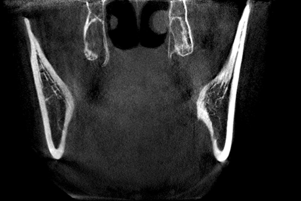
A coronal slice cuts across the arch like a slice of bread cut from the front. Through the molars it shows both sides of the mandible in the same picture: the alveolar crest, the thickness of the buccal and lingual plates, the roots of a molar in the bone, and the canal as a round dark spot low in the body on each side. Where the maxilla is included, the coronal plane is the classic view for the floor of the maxillary sinus and the floor of the nose above the roots.
Coronal is the plane for comparing sides. A difference in bone height, cortical thickness or the position of a canal is obvious when left and right sit next to each other on one slice. It is viewed as if facing the patient, so again the patient's left is on your right.
Sagittal: the profile view
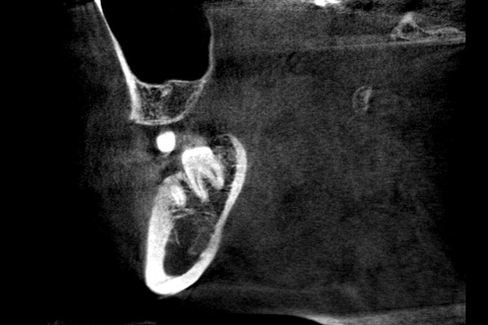
A sagittal slice runs from front to back and shows the jaw in profile. A slice through the molar region shows a tooth from crown to apex with the bone around it, the inclination of the roots, and the mandibular canal running forward and down towards the mental foramen. A midline sagittal slice shows the symphysis and the lingual concavity below the incisors, which is hard to appreciate in any other plane.
In JawAtlas a sagittal slice is displayed with anterior on the viewer's left. Because the dental arch is curved, a sagittal slice is only perpendicular to the arch near the midline. In the molar region it cuts the arch obliquely, which is why cross-sections perpendicular to the traced arch are used instead when the buccal to lingual width of the bone matters. See the panoramic reconstruction guide for how those are made.
How a slice stack is built from voxels
The volume is a three dimensional grid of voxels, each holding one grey value. A slice in any of the three standard planes is one layer of that grid: all the voxels that share the same index along one axis. An axial slice takes one layer along the head to feet axis, a coronal slice one layer along the front to back axis, and a sagittal slice one layer along the left to right axis.
Because the standard planes align with the grid, no interpolation is needed to build them, and when voxels are isotropic (the same size in every direction) all three stacks have the same resolution. The demo scan in JawAtlas has 480 x 480 x 320 voxels, so the axial stack has 320 slices and the coronal and sagittal stacks have 480 each. The field of view and voxel size guide works through those numbers. An oblique slice, such as the free Slice stage, cuts the grid at an angle and so does need interpolation between voxels.
The slice position readout
As you scroll, a readout at the top left of the pane shows the position of the current slice in millimetres, for example "Axial +10.2 mm". The number is the signed distance of the slice from the centre of the volume along the plane's normal. Zero is the middle slice; the sign tells you which side of the middle you are on, and the value tells you how far. Because it is in millimetres rather than a slice index, it means the same thing on scans with different voxel sizes.
This readout is a useful anchor when you describe a finding to someone else or save a moment: "the canal is clearest at Coronal +22 mm" tells another reader exactly where to look. A 20 mm scale ruler at the bottom left of the pane gives a sense of size without taking a measurement. For actual numbers use the Measure tool described in measuring length and angle.
Scrolling conventions
Every viewer lets you step through a stack, but the controls differ. JawAtlas uses a vertical slider on the right of the MPR pane; drag it to move through the slices of whichever plane is shown. Placing the crosshair in one plane also changes which slice is displayed in the other two, so scrolling and crosshair placement are two routes to the same result.
Whatever the control, a few habits transfer between viewers. Scroll the whole stack before stopping anywhere. Scroll slowly through regions that matter, because a thin structure such as a cortical plate or a canal wall can be missed between two fast steps. And after you stop, read the position readout and the edge labels again, since both change as you move. The reading routine puts these habits into a fixed order.
Questions students ask
What is the difference between axial, coronal and sagittal slices?
Axial slices are horizontal and show the dental arch as a horseshoe. Coronal slices run from ear to ear and show left and right sides together. Sagittal slices run from front to back and show the jaw in profile. All three are cut from the same volume.
Which plane is best for seeing the mandibular canal?
No single plane. Axial slices show the canal as a dark line inside the body, coronal slices show it as a round dark spot on each side, and sagittal slices follow its course forward and down. Cross-sections perpendicular to the arch show its buccal to lingual position most clearly.
Why do coronal and sagittal slices look as sharp as axial slices on CBCT?
Dental CBCT voxels are usually isotropic, the same size in every direction, so a layer taken along any axis of the grid has the same resolution. In medical CT with thick slices this is not the case, and reformats look blurred in one direction.
What does the millimetre number at the top of the slice mean?
It is the signed distance of the current slice from the centre of the volume along the plane's normal. Zero is the middle slice, the sign tells you which side of the middle you are on, and the value is in millimetres so it means the same on scans with different voxel sizes.
JawAtlas is an education and training aid. It is not a medical device and must not be used for diagnosis, treatment planning or any clinical decision.
Last reviewed 2026-10-13