 JawAtlas
Tri-planar MPR in CBCT, explained for dental students
What axial, coronal and sagittal views are, how the crosshair and scout lines link them, and how to read orientation labels on a dental CBCT scan.
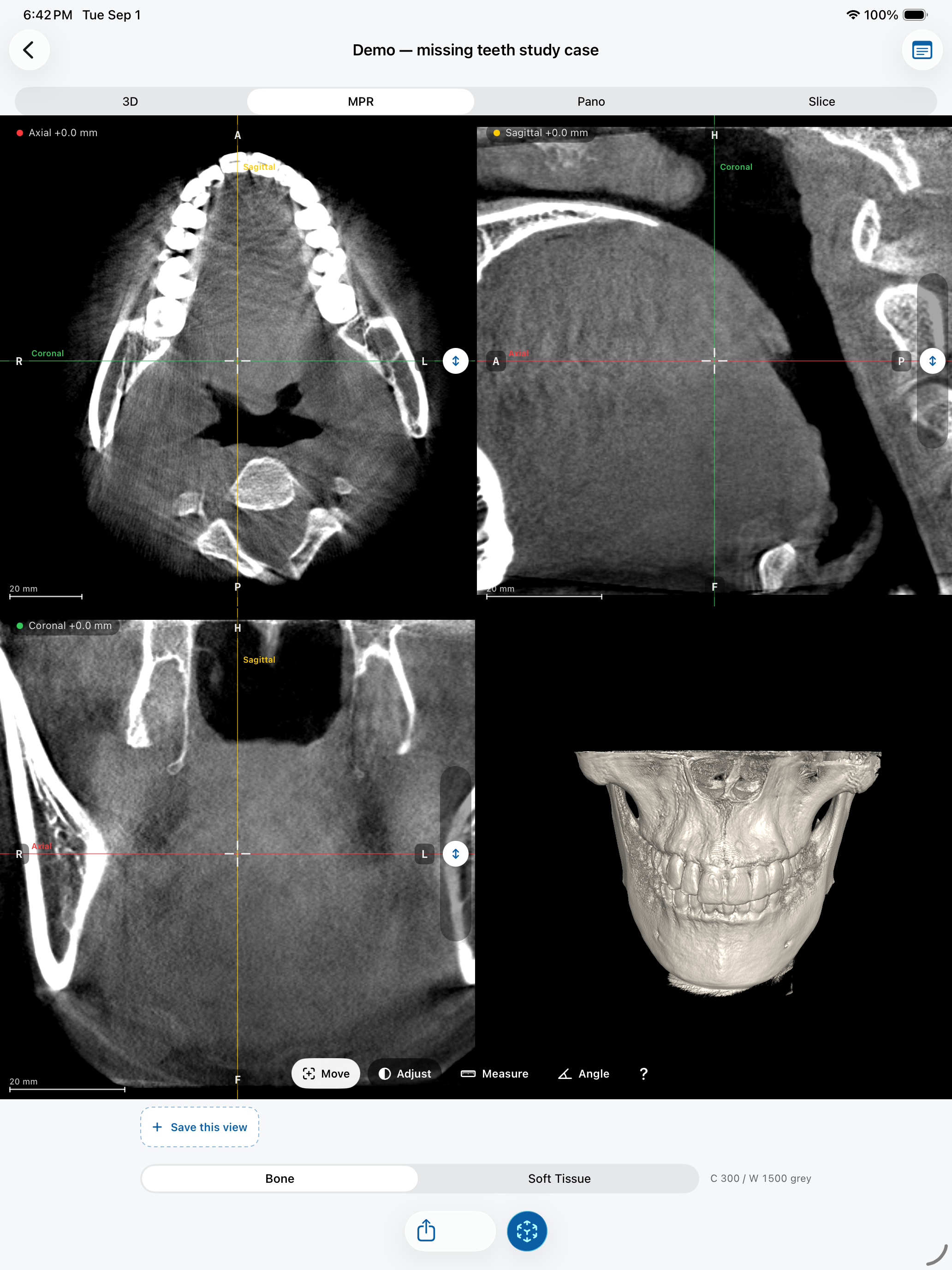
A CBCT scan is not a stack of pictures. It is a block of volume elements, or voxels, each holding a grey value. Multiplanar reconstruction (MPR) slices that block along three planes at right angles to each other, so you can look at the same point in the jaw from three directions at once.
The three planes
- Axial slices are horizontal, like looking down at the jaw from above or up from below. They are where you usually start: each slice shows the arch as a horseshoe.
- Coronal slices run from ear to ear, as if you were facing the patient. They show the relationship between left and right sides, the sinuses and the floor of the nose.
- Sagittal slices run from front to back, as if you were looking at the patient's profile.
Dental CBCT voxels are usually isotropic, meaning the same size in every direction. That is why a coronal or sagittal slice looks just as sharp as an axial one: no direction is reconstructed from coarser data.
The crosshair and scout lines
The three panes share one point in space, marked by a crosshair. Move it in any pane and the other two jump to the slices that pass through that point. The coloured scout lines in each pane show where the other two planes cut through it. Reading MPR well mostly comes down to this habit: find a structure in one plane, then confirm it in the other two before you believe what you see.
Reading orientation labels
Each pane has letters on its edges: R and L for the patient's right and left, A and P for anterior and posterior, H and F for head and feet. Radiology uses a fixed convention: axial slices are viewed as if from the patient's feet, and coronal slices as if facing the patient. In both, the patient's left appears on the right side of the screen. This surprises almost every beginner, so check the labels instead of assuming.
In JawAtlas the labels are calculated from the orientation stored in the scan itself, so they stay correct when the volume is rotated or cut at an oblique angle.
Common beginner mistakes
- Judging a finding from a single slice. A dark spot in one plane can be a canal, a foramen or a partial-volume effect; the other planes tell you which.
- Mixing up left and right on axial and coronal views. Read the labels.
- Measuring on a slice that does not pass through the widest part of the structure. Move the crosshair until it does, then measure.
Try it in JawAtlas
Open the bundled demo scan, switch to Move mode and drag the crosshair. Watch the scout lines follow in the other panes, then switch to Measure to take a length in millimetres.
Get JawAtlas free on the App StoreThis guide is for education and training. JawAtlas is not a medical device and must not be used for diagnosis, treatment planning or any clinical decision.