 JawAtlas
Guide
CBCT field of view and voxel size explained
After this guide you will be able to read the field of view and voxel size of any CBCT scan, say what they mean for the structures you can see, and work through the numbers on the demo scan yourself.
What field of view means
The field of view (FOV) is the volume of anatomy the scanner actually captures. On a CBCT it is usually a cylinder, described by its diameter and height in centimetres, and sometimes a sphere. Everything inside the cylinder is reconstructed; everything outside it is not in the scan at all, though it can still affect the image by scattering radiation into the detector.
Manufacturers group fields of view into rough sizes. A small field, about 4 to 6 cm across, covers a few teeth and the bone around them in one jaw. A medium field, around 8 to 12 cm, covers one full arch or both jaws together. A large field, 15 cm and more, covers the jaws, the sinuses and much of the skull. The exact boundaries vary by machine and these are plain language groupings, not standards. In general a larger field covers more anatomy and a smaller field allows a finer voxel, because the detector's pixels are spread over less tissue.
What a voxel is and what resolution means
A CBCT volume is a grid of voxels, the three dimensional equivalent of pixels. Each voxel is a small box of tissue represented by a single grey value. The voxel size is the edge length of that box, and dental CBCT voxels are typically a few tenths of a millimetre. The planes guide explains how slices are cut from this grid.
Voxel size sets the sampling grid, and nothing smaller than the grid can be represented. A structure thinner than one voxel, such as a very thin cortical plate or a periodontal ligament space, is averaged with its neighbours and may fade or vanish. This is the partial volume effect, and it is a property of the grid rather than of the anatomy. But voxel size is not the same as resolution. The sharpness you see also depends on the scanner's focal spot, the detector, the reconstruction filter, patient movement during the sweep and noise. A very small voxel on a noisy scan can look worse than a slightly larger one, and two scanners with the same voxel size can produce quite different images.
The demo scan as a worked example
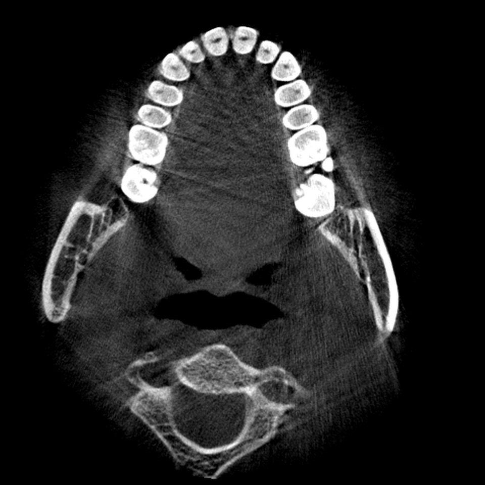
The scan bundled with JawAtlas is a good place to practice reading these numbers. Its volume is 480 x 480 x 320 voxels. The pixel spacing within a slice is 0.266 x 0.266 mm and the spacing between slices is 0.252 mm. Multiply and you get the physical size of the field: 480 x 0.266 mm is about 127.7 mm across in each in-plane direction, and 320 x 0.252 mm is about 80.6 mm in height, so roughly 127 x 127 x 80 mm. On the manufacturer scale above that is a large field for a mandible: it includes the full arch, both rami and a traceable inferior alveolar canal.
Count the slices, too. The axial stack has 320 slices, and the coronal and sagittal stacks have 480 each, because those planes cut across the 480 voxel in-plane axes. Scrolling from one end of the axial stack to the other therefore moves 80 mm through the jaw in 320 steps of a quarter of a millimetre. A molar root about 12 mm long spans roughly 48 axial slices, which is why a fast pass can skip the apex entirely and a slow pass is part of the reading routine.
| Quantity | Demo scan value | How it is derived |
|---|---|---|
| Voxel grid | 480 x 480 x 320 | Rows, columns and number of slices from the DICOM tags |
| Voxel size | 0.266 x 0.266 x 0.252 mm | Pixel Spacing and the spacing between slice positions |
| Field of view | about 127 x 127 x 80 mm | Grid size multiplied by voxel size on each axis |
| Axial slices | 320 | One per layer along the slice normal |
| Coronal and sagittal slices | 480 each | One per layer along each in-plane axis |
Isotropic versus anisotropic voxels
A voxel is isotropic when it has the same size in all three directions and anisotropic when it does not. Most CBCT scanners reconstruct isotropic or nearly isotropic voxels, which is one of the format's advantages over older medical CT with thick slices. The demo scan is very close: 0.266 mm in plane and 0.252 mm between slices, a difference of about five percent that is invisible in practice.
Anisotropy matters most when the spacing between slices is much larger than the in-plane spacing, for example 0.3 mm pixels with 1 mm between slices. The axial slices still look fine, but a coronal or sagittal reformat samples only one voxel per millimetre in the vertical direction, so it looks stepped or stretched and small structures that cross the slices are blurred. When you look at a reformat from a scan you did not make, check the spacing tags before trusting fine vertical detail. The DICOM guide explains where those tags live.
Why slice spacing matters for reformats
Everything in the MPR, Pano and Slice stages is a reformat, a picture computed from the volume by sampling it along some plane or curve. A standard axial, coronal or sagittal slice samples one layer of the grid and needs no interpolation. A free oblique slice, a panoramic reconstruction along a traced arch, or a cross-section perpendicular to that arch cuts the grid at an angle and must interpolate between neighbouring voxels. The accuracy of that interpolation depends on how far apart the voxels are in every direction, and the largest spacing sets the limit.
Measurement follows the same rule. The Measure and Angle tools in JawAtlas are calibrated from the DICOM spacing, so a distance read on an oblique or panoramic view is correct in millimetres only as far as the volume is correctly sampled. A length taken along the stack on an anisotropic scan is more uncertain than one taken within a slice. With isotropic voxels the uncertainty is the same in every direction, which is why near-isotropic data makes the panoramic reconstruction and the cross-sections in the panoramic guide trustworthy, and why the whole JawAtlas volume can live in a single 3D texture with one sampling rule. For how measurements are taken and read, see measuring length and angle.
Questions students ask
What does field of view mean on a CBCT scan?
The field of view is the size of the cylinder or sphere of anatomy the scanner captures, usually given as diameter by height in centimetres. A small field covers a few teeth, a medium field one or both jaws, and a large field most of the skull.
What is a voxel?
A voxel is the smallest element of a volume, the three dimensional equivalent of a pixel. Each one holds a single grey value for a small box of tissue. The size of that box, for example 0.25 mm on each side, is the voxel size.
Does a smaller voxel always mean a sharper image?
Not on its own. A smaller voxel sets a finer sampling grid, but the sharpness you see also depends on the scanner's focal spot, the detector, patient movement and noise. A very small voxel on a noisy scan can look worse than a slightly larger one.
What is the difference between isotropic and anisotropic voxels?
Isotropic voxels are the same size in all three directions, so slices in any plane have the same resolution. Anisotropic voxels are longer in one direction, usually between slices, so reformats in that direction look stretched or stepped.
How many slices are in the JawAtlas demo scan?
The volume is 480 x 480 x 320 voxels, so there are 320 axial slices, each 480 by 480 pixels, and 480 slices in each of the coronal and sagittal stacks. At 0.266 mm in plane and 0.252 mm between slices that covers about 127 x 127 x 80 mm.
JawAtlas is an education and training aid. It is not a medical device and must not be used for diagnosis, treatment planning or any clinical decision.
Last reviewed 2026-10-13