 JawAtlas
Guide
CBCT artifacts and how to recognise them
After this page you will be able to name the common artifacts on a dental CBCT scan, say what causes each one, tell them apart from anatomy, and predict how they change when you move the window.
What an artifact is
An artifact is anything in the image that is not in the patient. CBCT acquires a few hundred two dimensional projections while the tube and detector swing around the head, then reconstructs a three dimensional block of voxels from them. Every step of that process makes assumptions: that the X-ray beam has a fixed energy, that the head did not move, that the detector responds evenly, that each voxel holds one material. Where an assumption fails, the reconstruction puts something into the image that was never there.
Learning to read CBCT is partly learning to see past artifacts, so that you neither mistake a streak for a fracture line nor dismiss a real finding as noise.
Beam hardening and streaks from metal
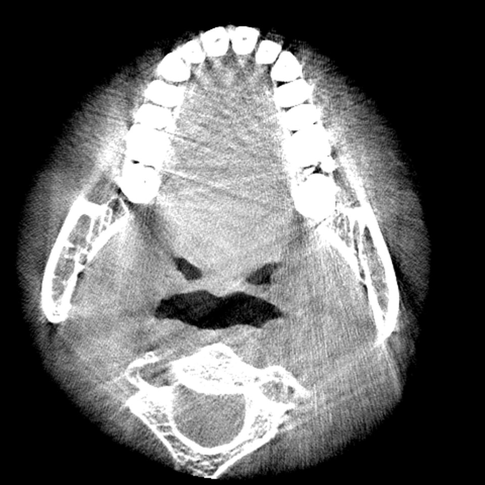
An X-ray beam contains a range of photon energies. As it passes through dense material the low energy photons are absorbed first, so the beam that comes out is on average more energetic, or harder, than the beam that went in. The reconstruction assumes a single energy, and the mismatch shows as dark bands between dense objects and bright streaks radiating from them.
Metal restorations, crowns, posts, implants and orthodontic brackets are the strongest sources. Around them you see a starburst of bright lines and dark shadows, and the bone between two metal objects can go almost black. Even tooth enamel, the densest natural tissue, causes a mild version: on an axial slice through the crowns, faint dark bands run between neighbouring teeth. Two clues separate artifact from anatomy: streaks are straight, radiate from a dense object, and cross anatomical boundaries.
Motion artifact
A CBCT scan takes several seconds, and a patient who moves during it is imaged in two positions at once. The reconstruction cannot reconcile the projections, so edges double, cortical outlines look smeared, and fine structure such as trabecular bone turns into a blur. The most reliable sign is a second faint outline running parallel to a cortical plate or a tooth, like a ghost edge.
Motion affects the whole volume, so scroll through the stack and look at distant structures before you decide. A doubled outline on both rami is movement; blur in one small area is more likely something else.
Noise
Noise is random grain. Every voxel's grey value includes a random error that depends on how many photons reached the detector, so thinner patients, lower exposure settings and smaller voxels all give grainier images. Noise is uniform and has no direction, which is how you tell it from streaks. It is most visible in soft tissue, where the true grey values are close together, and it is why a soft tissue window on CBCT often looks rough compared with a medical CT.
Partial volume effect
A voxel holds one number, the average of everything inside it. Where a structure is thinner than a voxel, or where a boundary runs obliquely through the voxel, that number is a blend of two materials. The result is that thin bright structures such as the lamina dura, a thin cortical plate or the rim of the inferior alveolar canal look fainter than they are, or vanish on one slice and reappear on the next. A dark space can look as if its wall is missing when the wall is simply thinner than the voxel.
The effect is worse with larger voxels, which is one reason voxel size matters when choosing a scan protocol (see field of view and voxel size). The remedy when reading is to look in the plane that cuts the structure at right angles: a cortical plate seen edge on is thin, but a plate seen obliquely is smeared across several voxels and seems thicker and fainter.
Ring artifacts
If one detector element reads slightly differently from its neighbours, that error is present in every projection at the same place. Reconstructed, it becomes a circle or arc centred on the axis of rotation. Ring artifacts are easiest to see on axial slices as faint concentric circles. They come from the machine, not the patient, ignore anatomy and are perfectly round, so they are easy to identify once you have seen one.
Scatter
Some photons are deflected inside the patient and land on the detector in the wrong place. The flat panel in a CBCT unit catches a large cone of X-rays at once, so it receives much more scatter than a medical CT with its narrow fan beam. Scatter adds a smooth haze to the projections and lowers contrast. In the volume it shows as a general greyness, a brighter edge and darker centre in large objects (called cupping), and a background that drifts across the scan. This drift is one reason CBCT grey values are not calibrated Hounsfield units: the same bone can read differently depending on where it sits in the field of view (see window, level and grey values).
How windowing changes their appearance
Windowing cannot remove an artifact, because the artifact is in the voxel data. What it changes is how visible the artifact is. A narrow window stretches a small range of grey values across the whole brightness scale, so streaks, noise and cupping all become stronger. A wide window compresses them and the image looks cleaner, at the price of less contrast everywhere.
If you suspect a faint line is a streak, narrow the window: a streak gets more obvious and stays straight, while a real thin cortical line keeps its anatomical shape. If you suspect noise, widen the window and see whether the texture smooths out. In JawAtlas, switch between the Bone and Soft Tissue presets on the same slice and then fine tune in Adjust mode, and watch what changes (see adjusting window and level).
Why artifacts matter when you learn to read
Artifacts matter for three reasons. They hide things: the bone around a metal crown is often unreadable, and motion can erase every fine detail. They imitate things: a dark beam hardening band between two roots can look like a gap in bone, and partial volume can make a thin wall look absent. And they shape habits. A student who checks for artifacts before describing anatomy learns to ask where the metal is, whether the patient moved, and which plane cuts a structure at right angles, the same questions that make every other part of reading more reliable (see how to read a CBCT scan).
Questions students ask
Why are there dark and bright streaks around fillings and crowns on CBCT?
Metal absorbs the lower energy part of the X-ray beam, so the beam that passes through it is harder than the scanner expects. The reconstruction turns that into dark bands and bright streaks that radiate from the metal, called beam hardening or metal artifact.
How can I tell motion artifact from a blurry scan?
Motion gives double contours and a smeared, misregistered look, often with a second faint outline of the cortical bone or teeth. Look at the whole volume: if the blur is everywhere and in all directions it is more likely noise or low resolution, but doubled edges point to movement during the scan.
What is the partial volume effect?
Each voxel holds one grey value that averages everything inside it. Where a thin structure such as the lamina dura or a cortical plate is thinner than a voxel, its value is averaged with its neighbours, so it looks fainter or seems to vanish. Larger voxels make the effect worse.
Does changing the window remove artifacts?
No. Windowing only changes which grey values are shown bright and dark. A narrow window makes streaks and noise more obvious, a wide one hides them, but the artifact is still in the data.
JawAtlas is an education and training aid. It is not a medical device and must not be used for diagnosis, treatment planning or any clinical decision.
Last reviewed 2026-10-13