 JawAtlas
Guide
Maximum intensity projection (MIP) on a dental CBCT
After this guide you will know what a maximum intensity projection is, why it looks like a radiograph, what it throws away, and when to switch to it in the 3D stage instead of a shaded rendering.
What a MIP is
A maximum intensity projection is one of the simplest ways to turn a volume into a picture. Imagine the volume as a block and the screen as a window in front of it. For every pixel of the window, the renderer sends a straight ray through the block and looks at every voxel the ray passes. It keeps the single highest grey value it finds and paints the pixel with that value. Everything else along the ray is thrown away.
Because the rule is "keep the brightest", the densest structures win. On a dental CBCT that means enamel, then cortical bone, then dentine and dense trabecular bone. Soft tissue almost never wins a ray, because somewhere along most rays there is bone or tooth that is brighter. The result is a picture in which teeth and the cortical outline of the jaw stand out against a dark background, with no surfaces and no lighting.
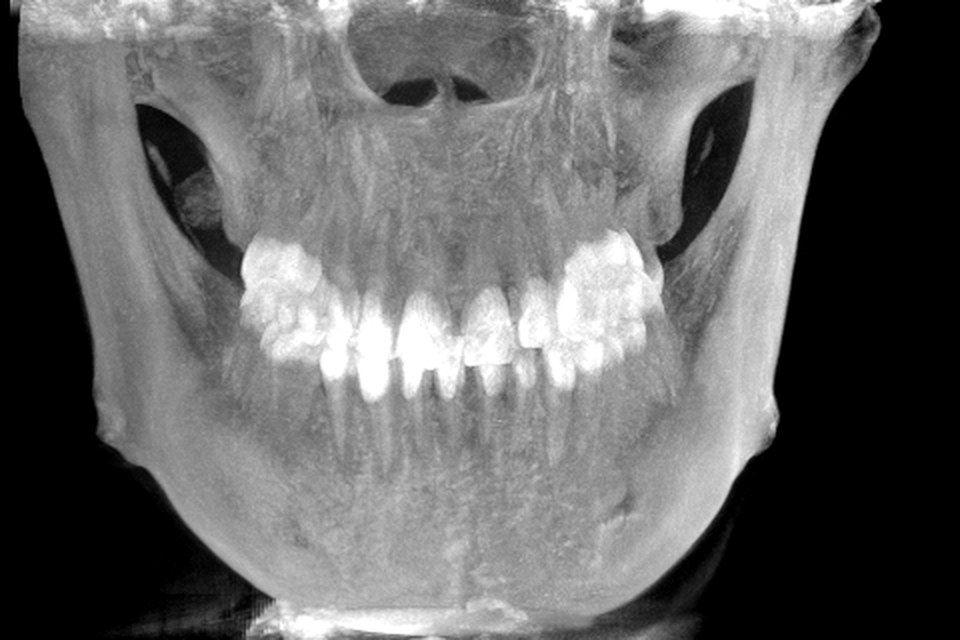
Why it reads like a radiograph
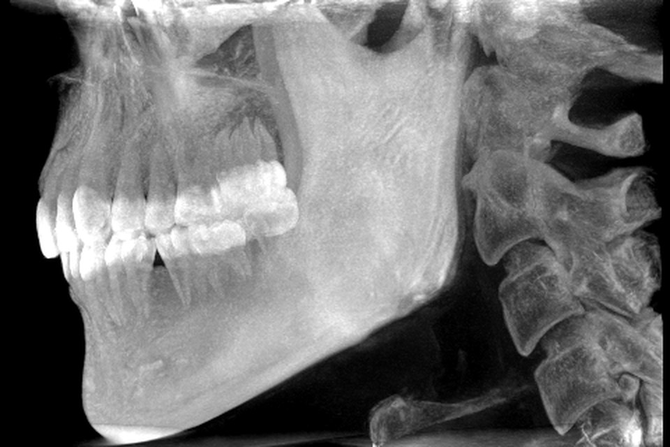
A radiograph is also a projection: X-rays cross the whole patient and the detector records how much got through along each path. Structures at every depth are superimposed on the film. A MIP superimposes in the same way, which is why a frontal MIP of a jaw looks like a posteroanterior radiograph and a lateral MIP looks like a lateral skull view. Students who have read plain films find a MIP immediately familiar.
The two are not identical. A radiograph sums attenuation along the ray, so thick soft tissue adds a grey haze and two thin dense objects in a line look denser than one. A MIP keeps only the maximum, so soft tissue vanishes, contrast is harsher, and overlapping dense objects look no brighter than one. Roots within bone are often clearer on a MIP than on a film for this reason, while subtle density differences are lost.
What a MIP hides: depth
The price of a projection is that depth disappears. Two structures at different distances along the same ray land on the same pixel, and nothing in the picture tells you which is nearer. A MIP also discards everything less dense than the winner on its ray. The mandibular canal is a dark channel inside dense bone; on a MIP the bone wins every ray that crosses the canal, so the canal is simply gone. The same is true of the pulp chamber, the periodontal ligament space and any radiolucency surrounded by cortex.
This is why a MIP is a view for overview and for dense structures, not for reading anatomy inside bone. If you rotate a MIP the superimposition changes and you can sometimes infer which structure lies in front, but the slices remain the only place where depth is explicit. The reading routine puts every projection and rendering after the slice passes for this reason.
MIP versus shaded surface rendering
A shaded rendering answers a different question. Instead of "what is the densest thing on this ray", it asks "where does this ray first meet a surface", using a threshold or a transfer function to decide what counts as surface. It then lights that surface with a virtual lamp so that near things look different from far things and curved things show their shape. The result is the familiar solid jaw that can be rotated like a model. Depth is restored, but only for the surfaces the threshold selected; anything inside or below them is hidden, and anything less dense than the threshold does not exist in the picture.
So the two renderings fail in opposite ways. A MIP shows dense structures at every depth but cannot say which is in front. A shaded surface says exactly what is in front but shows nothing behind it. A thin cortical plate that falls below the threshold vanishes from a shaded rendering but may still show on a MIP, because somewhere along its ray a voxel is bright enough. Used together, each corrects the other.
| Property | MIP | Shaded surface rendering |
|---|---|---|
| Rule per ray | Keep the brightest voxel | Stop at the first surface and shade it |
| Depth cues | None | Lighting and occlusion |
| Looks like | A radiograph | A solid model |
| Hides | Everything less dense on the ray | Everything behind or below the surface |
| Good for | Overview of teeth and cortical outline | Shape and spatial relationships of surfaces |
Using MIP mode in the 3D stage
In JawAtlas the 3D stage renders the volume by ray marching on the GPU. The Bone, Teeth and Soft tissue presets choose which grey values are shown and how, and the Shaded toggle adds lighting. Tap MIP to switch to maximum intensity projection; the picture changes from a lit surface to a radiograph-like projection of the same volume. Drag to rotate, pinch to zoom, and double-tap to reset the view.
A useful exercise is to rotate slowly in MIP mode from a frontal view to a lateral one and watch structures slide across each other as the superimposition changes. Then switch MIP off, keep the same angle, and see what the shaded rendering adds and removes. Finish by going to the MPR stage and finding the structure you were watching in the three planes, where its depth is no longer a guess. The 3D volume rendering feature page lists the controls, and the grey values guide explains why none of these presets measure density.
Questions students ask
What is a maximum intensity projection?
A MIP is a 2D image made by sending a ray through the volume for each pixel and keeping only the brightest voxel the ray meets. Dense structures such as enamel and cortical bone dominate, and everything less dense along that ray is discarded.
Why does a MIP look like an X-ray?
Both are projections of the whole volume onto a plane, so dense structures from every depth are superimposed and the picture resembles a radiograph. A radiograph sums attenuation along the ray while a MIP keeps the maximum, so a MIP has stronger contrast and no soft tissue haze.
What does a MIP hide?
Depth. Two structures at different distances along the same ray land on the same pixel and you cannot tell which is in front. A MIP also hides anything less dense than the brightest voxel on its ray, so a canal inside dense bone disappears.
What is the difference between MIP and shaded surface rendering?
A shaded rendering decides where a surface is, lights it, and draws the first surface the ray hits with depth cues. A MIP ignores surfaces and lighting and shows the densest voxel on every ray regardless of depth. Shaded views look solid; MIPs look like radiographs.
JawAtlas is an education and training aid. It is not a medical device and must not be used for diagnosis, treatment planning or any clinical decision.
Last reviewed 2026-10-13