 JawAtlas
Guide
CBCT versus a panoramic radiograph: what each image shows
After this guide you will be able to explain how a conventional panoramic radiograph is made, how a panoramic reconstruction from a CBCT volume differs from it, and why each one is taught.
What a panoramic radiograph is
A panoramic radiograph, often called an OPG or orthopantomogram, is a single two dimensional image of both jaws from condyle to condyle. It is made during the exposure, not afterwards. An X-ray tube and a detector rotate around the patient's head on opposite sides while the detector records a narrow strip at a time. The centre of rotation moves during the sweep so that one curved layer, the focal trough, stays sharp while everything in front of or behind it is blurred out.
The result is a projection image. Every pixel sums the attenuation of all the tissue the beam crossed on its way to that part of the detector, within the layer that was kept in focus. There is no depth information: a structure cheek side of the arch and one tongue side of it land on the same spot in the picture. This is the image most dental students learn to read first, because it is quick, low in dose compared with CBCT, and shows the whole dentition in one glance.
How a panoramic reconstruction from CBCT is made
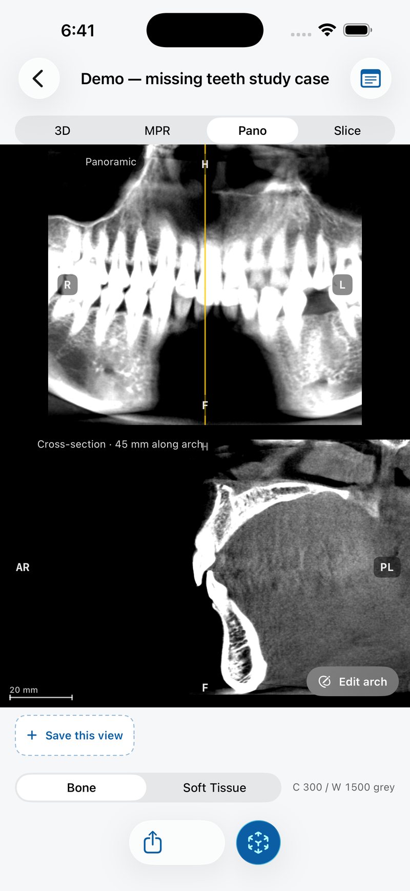
A CBCT scan does not produce a panoramic image at all. It produces a volume of voxels, from which a viewer can compute a panoramic view after the scan. You trace a curve along the dental arch on an axial slice, the software builds a smooth path through your points, and then it samples the volume along short lines that cross the curve at right angles. Each column of the resulting image comes from one of those lines, so the arch is unrolled flat.
In JawAtlas this happens in the Pano stage. The demo scan ships with a default arch; tap "Edit arch" to place at least four points of your own along the alveolar process, then "Show panoramic" to rebuild. The view is computed on the GPU by sampling along the curve with an averaged slab. The panoramic reconstruction guide walks through the method step by step and the arch tracing how-to shows the taps.
Three ways the two images differ
Slab thickness
An OPG has a focal trough whose thickness is set by the machine and varies around the arch: wider at the molars, narrower at the incisors. A CBCT pano has a slab whose thickness is chosen in software. The slab is the distance on either side of the curve over which voxels are averaged. A thick slab looks smooth and familiar; a thin slab is sharp but shows only what lies very close to the curve you drew. Either way, the slab is explicit and the same everywhere along the arch, which the focal trough is not.
Magnification and distortion
In an OPG the beam diverges from the tube, the detector moves, and the centre of rotation shifts, so horizontal and vertical magnification differ from each other and change with position in the arch. Objects placed slightly outside the trough are stretched or compressed, and structures on the opposite side of the head, such as the contralateral ramus or the hard palate, cast blurred ghost images. A CBCT pano is sampled from a volume whose voxel size is known in millimetres, so there is no projection magnification and no ghosting. Distances along the curve are true to the volume, which is why the Measure tool in JawAtlas can read millimetres on the pano and the cross-section alike.
The traced arch
An OPG's focal trough is fixed by the machine and the operator's positioning. A CBCT pano depends on the curve you trace, and two people can produce quite different pictures from the same scan. A curve that drifts buccal or lingual makes teeth look narrower, broader or blurred. This is a weakness, because it adds a human step, and a strength, because you can retrace and rebuild in seconds. On a CBCT the pano is a reformat you control, not a fixed image you receive.
Where each belongs in teaching
The OPG is taught early because it is the most common extraoral image in general practice and because reading it well is a skill of its own: recognising the normal anatomy, the artifacts of positioning, the ghost images and the limits of a projection. Students who skip it and go straight to CBCT miss the reasoning that explains why a projection can mislead.
CBCT is taught for the questions a projection cannot answer: where a structure sits in the buccal to lingual dimension, how thick the bone is at a point, how a root and a canal relate in three dimensions. A reconstructed pano is the bridge between the two. It shows the familiar overview, and the scout line drags a perpendicular cross-section through the jaw so that the hidden dimension is one tap away. Moving between the pano and the cross-section is the best way to understand what a flat image leaves out. The reading routine places this step after the three planes and before 3D.
| Property | Panoramic radiograph (OPG) | Panoramic reconstruction from CBCT |
|---|---|---|
| How it is made | Projection during exposure, rotating tube and detector | Computed after the scan from the volume |
| Sharp layer | Focal trough set by the machine | Slab around a curve traced by the user |
| Magnification | Uneven, varies by position | None in the projection sense; voxel size is known |
| Ghost images | Present from contralateral structures | Absent |
| Depth information | None | Available through cross-sections |
| Dependence on the operator | Patient positioning | Arch tracing and slab choice |
Limits of both
An OPG cannot separate structures that overlap along the beam, cannot show bone width, and distorts anything outside its trough. Reading it demands constant awareness of what a projection does to anatomy.
A CBCT pano has limits too. It only shows what lies within the slab around the curve, so a root that curves out of the slab fades out even though it is present in the volume. A poorly traced arch produces a misleading picture with no warning. Metal restorations produce streaks that run through the slab, and the grey values are not calibrated, so density cannot be read from the image; see the artifacts guide and the grey values guide. The cross-section fixes the depth problem but only at one position at a time, which is why the routine is to step it along the arch rather than to look once.
Finally, neither image is a substitute for the other in clinical practice, and this site does not advise on which to take. For learning, the useful exercise is to look at the same tooth in a reconstructed pano, in its cross-section, and in the three standard planes, and to work out why each picture looks the way it does.
Questions students ask
Is a panoramic reconstruction from CBCT the same as a panoramic radiograph?
No. An OPG is a projection made during exposure by a rotating tube and detector that keep one curved layer sharp. A CBCT pano is computed afterwards from the volume by sampling a slab along a curve you trace. They look alike but are produced in different ways and have different limits.
Why does a panoramic radiograph magnify and distort the image?
The beam diverges from the tube and the focal trough moves around the arch, so horizontal and vertical magnification differ and change with position. Objects outside the trough blur, and structures on the opposite side cast ghost images. A CBCT pano samples a measured volume, so it has none of these projection effects.
What does slab thickness do in a CBCT panoramic reconstruction?
The software averages voxels across a short distance on either side of the traced curve. A thicker slab looks smoother and more like an OPG but blurs small detail; a thinner slab is sharper but noisier and more sensitive to where the curve was drawn.
Why do dental schools still teach panoramic radiographs if CBCT exists?
The OPG remains an everyday image with its own reading skills, lower dose and wide availability. CBCT gives three dimensional information at a higher dose and cost. Students need to read both and to understand what each cannot show.
JawAtlas is an education and training aid. It is not a medical device and must not be used for diagnosis, treatment planning or any clinical decision.
Last reviewed 2026-10-13