 JawAtlas
Anatomy
The maxillary sinus on CBCT
After this page you will be able to find the floor of the maxillary sinus on CBCT, see how it relates to the molar roots, recognise septa and the ostium, and know what the lining looks like in a soft tissue window.
What the sinus is
The maxillary sinus is the largest of the paranasal sinuses, a roughly pyramid shaped air space inside the body of the maxilla on each side. Its base is the lateral wall of the nasal cavity, its roof is the floor of the orbit, and its floor is the alveolar process above the premolars and molars. Air has the lowest grey values in the scan, so the sinus appears almost black, outlined by the thin bright cortical bone of its walls.
Because air and bone sit at opposite ends of the grey scale, the walls are easy to see. The difficult part is the floor, where thin bone, tooth roots and the lining meet.
The sinus floor and the molar roots
The floor is usually lowest around the first and second molars and rises toward the premolars and the third molar. The relationship between the floor and the roots varies a lot from person to person and even from tooth to tooth. A root may end well below the floor with thick bone between, sit against the floor with only a thin plate of bone, or bulge up into the sinus so that the floor drapes over it like a tent.
Coronal slices show this best: you see the root apex and the floor above it in one picture, with the nasal floor beside it for comparison. Sagittal slices show the whole row of roots against the undulating floor, which is the view that matches a panoramic radiograph. Axial slices at the level of the floor show which root tips reach that height, each one appearing as a small bright circle surrounded by bone or by dark air.
Septa
A septum is a ridge or partition of bone that rises from the floor or a wall of the sinus, partly dividing it. Septa are common, especially in the molar region. On a coronal or sagittal slice a septum looks like a bright spike or shelf standing up from the floor into the dark air space; on an axial slice above the floor it can look like a wall across the sinus.
On a single slice a septum can be mistaken for the floor itself, or for a root. Put the crosshair on it and look at the other two planes: a septum is continuous with the sinus wall, a root with a crown below (see how the three planes link).
The ostium in plain terms
The sinus drains into the nose through a small opening called the ostium, which sits high on the medial wall, near the roof, and opens into the middle meatus of the nasal cavity. In plain terms, the drain is near the top of the box, not the bottom. The ostium is best seen on a coronal slice, where it shows as a short dark channel between the sinus and the nasal cavity.
On many dental scans the ostium is above the field of view, because the scan was centred on the teeth. If you cannot find it, check the top edge of the volume first before assuming you have missed it.
How it looks in each plane
- Axial. A dark oval behind the cheek, medial to the zygoma. Scroll down and the oval shrinks until the floor closes it off; scroll up and it widens toward the orbit.
- Coronal. The classic textbook view: a dark triangle beside the nasal cavity, with the floor of the sinus usually lower than the floor of the nose in adults. The medial wall, the ostium and the roots below the floor are all in this plane.
- Sagittal. A dark box above the roots from the canine region back to the tuberosity. This plane shows the length of the sinus from front to back and how the floor steps up and down over each root.
Seeing the lining in a soft tissue window
In the Bone preset the sinus is simply black. Switch to the Soft Tissue preset and the grey values that sit between air and bone are spread across the brightness range, so anything of soft tissue density inside the sinus becomes visible as a mid grey layer between the black air and the white wall.
A healthy lining is too thin to show on CBCT. When a band of soft tissue grey value does line the floor or a wall, textbooks call the appearance mucosal thickening. You may also meet descriptions such as a dome shaped outline on the floor, or a flat upper surface of grey with black air above it. Learn to recognise these appearances and describe their position, extent and shape; what any of them means for a particular person is a clinical judgement, and JawAtlas is a learning tool, not a diagnostic one.
In Adjust mode you can refine the preset: drag up or down for level and left or right for width, and watch the lining appear and disappear as the window moves across its grey values. These are grey values, not Hounsfield units, so a window that works on one scan may need adjusting on another (see window, level and grey values).
What the demo scan shows, and what it does not
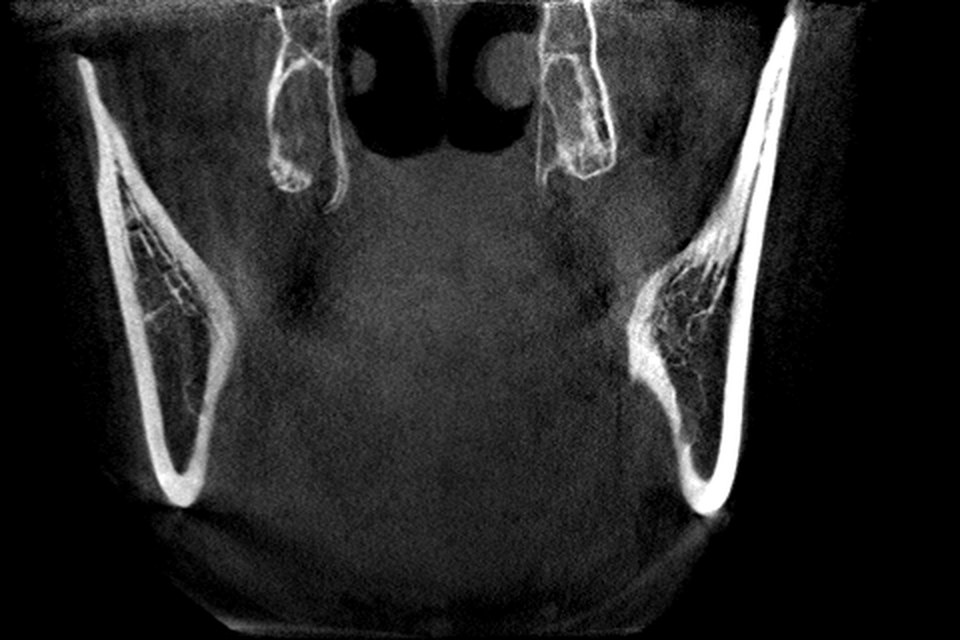
The bundled demo scan is a mandible-centred volume about 127 mm wide and 80 mm tall. The upper edge of the field of view cuts through the maxilla, so you see the upper teeth and the alveolar process, and at most the lowest part of the sinus floor on some slices. The rest of the sinus, the ostium and the orbit floor are outside the box. That is not a flaw: a mandible-centred field of view is a deliberate choice that trades coverage for a lower dose (see field of view and voxel size).
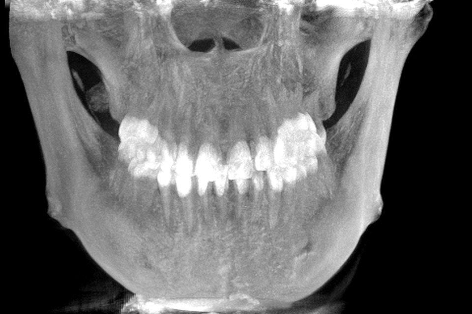
To study the full sinus you need a scan whose field of view includes the maxilla. Several openly licensed CBCT datasets do, and you can import a DICOM folder into JawAtlas through the Files app (see free CBCT datasets and how to import DICOM from Files). Then put the crosshair on the lowest point of the floor in the coronal plane and step through the sagittal stack to see how the floor rises and falls over each root.
Questions students ask
What does the maxillary sinus look like on CBCT?
A large, very dark air space inside the maxilla, above the premolar and molar roots, outlined by a thin bright wall of cortical bone. On an axial slice it is a dark oval, on a coronal slice a dark triangle beside the nasal cavity, and on a sagittal slice a dark box above the tooth roots.
Which plane shows the relationship between the sinus floor and the molar roots best?
Coronal, because it shows a root apex and the floor above it in the same picture, with the nasal cavity for comparison. Sagittal shows the whole row of roots against the undulating floor, and axial shows which roots are at the level of the floor.
What is mucosal thickening on a CBCT scan?
The name given to a band of soft tissue grey value lining the inside of the sinus wall, between the black air and the white bone. A normal lining is too thin to see. This page describes it only as an appearance to recognise; what it means in a particular person is a clinical question outside the scope of JawAtlas.
Why does the JawAtlas demo scan not show the whole sinus?
The demo scan is a mandible-centred field of view about 80 mm tall. Its upper edge cuts through the maxilla, so at most the lowest part of the sinus floor is inside the volume. To study the full sinus you need a scan whose field of view includes the maxilla.
JawAtlas is an education and training aid. It is not a medical device and must not be used for diagnosis, treatment planning or any clinical decision.
Last reviewed 2026-10-13