 JawAtlas
Anatomy
The incisive canal and nasal floor on CBCT
After this page you will be able to find the nasopalatine canal in the midline of the anterior maxilla, follow it from the incisive foramen to the nasal floor, and read the hard palate and nasal floor on sagittal and coronal slices.
What you are looking for
The nasopalatine canal, also called the incisive canal, runs through the midline of the anterior maxilla just behind the central incisors. Its lower end opens into the mouth at the incisive foramen, under the incisive papilla. Its upper end opens onto the floor of the nose, usually as two small openings either side of the nasal septum. It carries the nasopalatine nerve and vessels.
On CBCT it is a dark channel with a thin bright cortical wall, like the mandibular canal but shorter, wider at the bottom and running up and back rather than along the jaw. Its shape varies: a single tube, a Y with one oral opening and two nasal ones, or two parallel channels. Length and width differ from person to person, so learn the position and the course rather than a fixed size.
The sagittal slice: the classic view
A sagittal slice through the midline shows the whole canal at once, the textbook picture. Find the midline on an axial slice, put the crosshair between the two central incisors in Move mode and switch to sagittal. Scroll a slice or two either way with the slider until the dark channel appears. It rises from the incisive foramen on the palate side, just behind the root of the central incisor, and runs up and back to the nasal floor.
The same slice shows the hard palate as a bright plate of bone running back from the incisors, and above it the nasal floor, which is simply the upper surface of that plate. Below the palate is the dark air of the mouth or the mid grey of the tongue; above the nasal floor is the dark air of the nasal cavity.
The coronal slice
A coronal slice through the canal cuts it across, so you see a dark circle or oval in the midline between the roots of the central incisors, below the nasal floor. Scroll backwards through the stack and the circle moves up, following the slope of the canal. Near the nasal floor it often splits into two, one opening on each side of the septum.
Coronal is also the plane for understanding the palate. The nasal floor is a horizontal bright line with the two nasal cavities above it and the mouth below, and the nasal septum rises from its middle. Further back the maxillary sinuses sit either side of the nasal cavity, and in adults the sinus floor is usually lower than the nasal floor (see the maxillary sinus on CBCT).
The axial slice
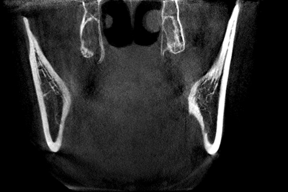
On an axial slice at the level of the upper incisor roots the canal is a dark oval in the midline immediately behind the central incisors. Scroll up and it narrows and moves backwards; just below the nasal floor it may become two small circles. Scroll down and it widens into the incisive foramen, where the palatal cortex opens into the mouth. The patient's left is on your right, and the midline is the line between the central incisors, not necessarily the centre of the picture (see orientation conventions).
Common confusions
A root for the canal. The central incisor roots sit either side of the canal, and on an oblique slice one can look like a dark channel because of its pulp. A root is mid grey with a thin dark pulp and a crown at one end; the canal is dark throughout with a bright rim.
A marrow space for the canal. The anterior maxilla has large marrow spaces. The canal is the one in the midline that connects the palate to the nasal floor; put the crosshair on it and check that it is continuous in the other two planes (see how the three planes link).
The nasal floor for the sinus floor. On a coronal slice behind the canines both are bright lines above the roots. The nasal floor is in the midline with the septum standing on it; the sinus floors are lateral and usually lower.
What the demo scan does and does not include
The bundled demo scan is a mandible-centred volume about 127 mm across and 80 mm tall. Its top edge passes through the maxilla a little above the nasal floor: the upper teeth, the incisive foramen and the hard palate are inside, the floor of the nasal cavity and the lowest part of the sinuses appear in the last slices, and anything higher is outside. So look for the canal near the top of the axial stack, and expect its nasal openings to sit close to the edge.
That is the field of view, not a fault in the scan (see field of view and voxel size). For the whole canal and the nasal floor you need a scan that includes the maxilla. Several openly licensed datasets do, and a DICOM folder imports through the Files app (see free CBCT datasets and how to import DICOM from Files).
A routine to practise
Start in the MPR stage with the Bone preset. On an axial slice at upper incisor root level, find the dark oval behind the central incisors and put the crosshair on it. Switch to sagittal and scroll until the channel runs up and back. Switch to coronal and step backwards through the stack, watching the circle climb towards the nasal floor. If the rim is faint, narrow the window width in Adjust mode; the numbers are grey values, not Hounsfield units, so the right setting differs between scans (see window, level and grey values). Save a moment at the foramen, mid-canal and at the nasal floor, and replay the three.
Questions students ask
What is the incisive canal on a CBCT scan?
The nasopalatine canal, a short channel in the midline of the anterior maxilla just behind the central incisors. It opens into the mouth at the incisive foramen in the palate and onto the nasal floor above, usually through two small openings either side of the septum. On CBCT it is a dark channel with a thin bright wall.
Which plane shows the nasopalatine canal best?
A sagittal slice through the midline shows its whole length, from the incisive foramen up and back to the nasal floor, with the hard palate in the same picture. Coronal and axial slices cut it across and are better for checking that a dark spot really is the canal and not a root or a marrow space.
Is the nasal floor the same as the hard palate?
They are the two surfaces of the same plate of bone. The hard palate is its lower, oral surface and the nasal floor its upper surface. On a coronal slice it is a horizontal bright line with the nasal cavities above and the mouth below.
Can I see the incisive canal on the JawAtlas demo scan?
Only its lower part, at best. The demo scan is centred on the mandible and its top edge cuts through the maxilla, so the incisive foramen region is near the top of the volume and the nasal floor is largely outside it. A scan that includes the maxilla shows the whole canal.
JawAtlas is an education and training aid. It is not a medical device and must not be used for diagnosis, treatment planning or any clinical decision.
Last reviewed 2026-10-14