 JawAtlas
Anatomy
The temporomandibular joint on CBCT
After this page you will be able to find the condyle, the glenoid fossa and the articular eminence on sagittal and coronal CBCT slices, explain why a slice set along the condylar axis matters, and know which scans actually include the joint.
The bony parts
The temporomandibular joint (TMJ) is where the condyle of the mandible meets the temporal bone. The condyle is the rounded head on top of the condylar process, which rises from the back of the ramus on a narrower neck. It sits in the glenoid fossa, a concavity on the underside of the temporal bone just in front of the ear canal. In front of the fossa the bone curves down and forward into a rounded ridge, the articular eminence, over which the condyle slides when the mouth opens wide.
Between condyle and fossa lie the articular disc and the joint capsule. These are soft tissue, which CBCT shows poorly, so on the scan they appear only as a dark band between the two bony surfaces. That band is the joint space, and on CBCT you study the bone on either side of it rather than the disc inside it.
The sagittal slice
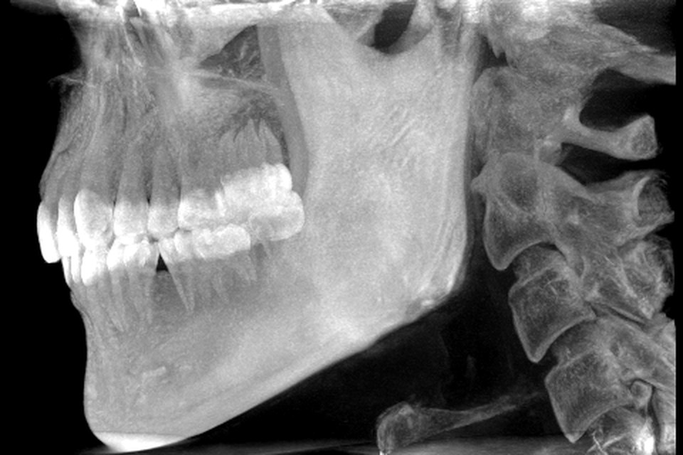
A sagittal slice through the condyle is the view most students know from textbooks. The condyle is a rounded cap with a thin bright cortical shell and grey trabecular bone inside, on top of the neck and ramus. Above it the fossa is a concave bright line, and its roof is a thin plate separating the joint from the cranial cavity. In front of the fossa the eminence bulges downwards. With the teeth together the condyle sits in the fossa; on a scan taken with the mouth open it has moved forward and down onto the eminence.
To get there in the MPR stage, find the condyle on an axial slice near the top of a scan that includes it, put the white crosshair on the centre of the head in Move mode and switch to sagittal. The slider then scrolls through the condyle from its lateral pole to its medial pole, and the slice position readout tells you how far you have moved (see how the three planes link).
The coronal slice
A coronal slice shows the condyle from the front: a rounded head, wider from side to side than the neck below it, with a medial pole and a lateral pole. The roof of the fossa arches over it as a thin bright line, with the dark joint space between. The medial pole usually sits a little lower than the lateral pole, and the neck leans inwards below the head. This is also the plane for comparing the two sides, because a coronal slice through both condyles shows their heights and shapes side by side.
Why a corrected sagittal plane matters
The two condyles are not lined up with the standard planes. On an axial slice through the heads each condyle is an elongated oval whose long axis runs backwards and inwards, so that the two axes, extended, would meet somewhere behind the skull base. The angle varies from person to person and from side to side.
A standard sagittal slice runs straight front to back, so it cuts the condyle obliquely. The head looks narrower or oddly shaped, the cortical shell can look thick on one side and missing on the other, and the joint space looks wider or narrower than it is. A corrected sagittal slice is one set perpendicular to the condyle's own long axis, and a corrected coronal slice is parallel to it. Both show the head, the space and the fossa in true profile, which is why textbook pictures are corrected cuts.
In JawAtlas the Slice stage is a free-oblique single slice that you can tilt in any direction, so on a scan that includes the joint you can bring a slice into line with the condylar axis by hand and compare it with the standard sagittal pane. Working out what to tilt, and which way, is itself good practice in thinking about the joint in three dimensions (see axial, coronal and sagittal planes).
Which scans include the joint
Whether you can study the TMJ depends entirely on the field of view. A large field of view scan, around 15 cm and more, covers both jaws, the sinuses and the skull base, so both joints are inside it with the condyles, fossae and eminences complete. A medium field of both jaws may catch the condyles at its upper edge or may not, depending on positioning. A small field centred on a few teeth never includes them (see field of view and voxel size).
The bundled demo scan is a mandible-centred volume about 127 mm across and 80 mm tall. Both rami run up to the top edge of the volume, and the condylar heads, the fossae and the eminences lie at or beyond that edge, so the joint cannot be studied there. What you can do with the demo scan is follow each ramus upwards on coronal and sagittal slices to the top edge and see how the neck leads towards the head. For the joint itself, import a large field of view case; some openly licensed datasets include the whole skull (see free CBCT datasets and how to import DICOM from Files).
A routine on a large field of view scan
Use the Bone preset, and narrow the window width in Adjust mode if the cortical shell looks faint. Scroll the axial stack up from the teeth until the ramus narrows into the neck and then widens into the oval head; stop there and note the direction of each head's long axis. Put the crosshair on the centre of one head and switch to coronal, then sagittal, saving a moment in each plane. Scroll through the whole head in sagittal, from lateral pole to medial pole, and watch the fossa and the eminence come and go above it. Repeat for the other side and replay the moments as a pair (see saving and replaying moments).
Questions students ask
What does the temporomandibular joint look like on CBCT?
On a sagittal slice the condyle is a rounded cap of bone on top of the ramus, sitting in the concave glenoid fossa with the rounded articular eminence in front. A dark band of soft tissue, the joint space, separates condyle and fossa. The disc itself is soft tissue and is not seen on CBCT.
Why do textbooks show corrected sagittal slices of the condyle?
The long axis of each condyle runs backwards and inwards, not straight across. A standard sagittal plane cuts it obliquely, which distorts its outline and the joint space. A slice set perpendicular to the condyle's own long axis shows the true shape and the fossa in true profile.
Can I see the TMJ on the JawAtlas demo scan?
No. The demo scan is a mandible-centred field of view about 80 mm tall. Both rami run up to the top edge of the volume, and the condylar heads, the fossae and the eminences lie at or beyond that edge. A large field of view scan is needed for the joint.
Does CBCT show the articular disc?
Not as a distinct structure. CBCT shows bone well and soft tissue poorly, so the disc and the joint capsule merge into the dark band of the joint space. Studying the disc needs a different kind of imaging; on CBCT you study the bony parts and the space between them.
JawAtlas is an education and training aid. It is not a medical device and must not be used for diagnosis, treatment planning or any clinical decision.
Last reviewed 2026-10-14