 JawAtlas
Anatomy
Finding the mental foramen on CBCT
After this page you will be able to find the mental foramen on a CBCT scan, recognise the anterior loop in front of it, and explain why the inferior alveolar canal seems to disappear just before it exits.
What you are looking for
The mental foramen is the opening on the buccal (outer) surface of the mandible through which the mental nerve and vessels leave the bone. It is the front door of the inferior alveolar canal, the structure you followed in finding the mandibular canal. Most often it sits below the second premolar or between the two premolars, roughly halfway between the alveolar crest and the lower border, but it can lie anywhere from the canine to the first molar.
On CBCT it is a gap in the bright buccal cortical plate with a dark channel behind it. The channel is the mental canal, the short final stretch that runs from the main canal up, out and back to the surface.
Start on the axial slice
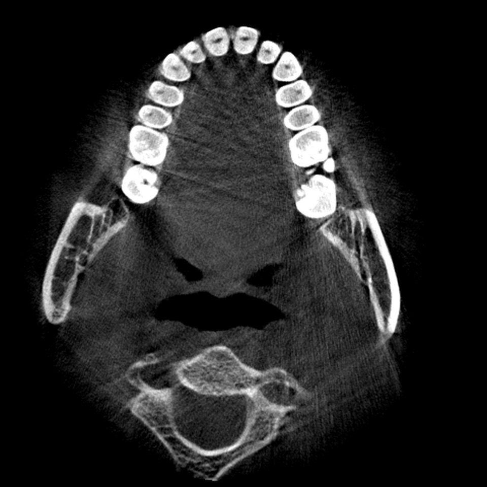
The axial plane is the easiest place to find it, because an axial slice cuts straight across the buccal cortex and shows the gap as a break in a white line. In the MPR stage choose Axial, start at the level of the premolar roots and scroll down slowly with the slider, watching the buccal cortex on each side. At some level a short dark notch opens in it and a dark channel curves in from behind. That is the foramen.
Scroll a few slices above and below. The notch usually shows on several consecutive slices, because the opening is a few millimetres tall and often faces up, back and out. Note the slice position readout so you can return to the level, and remember that the patient's left is on your right (see orientation conventions).
The anterior loop
In many people the canal does not simply turn out through the foramen. It runs forward past it, then curves up and back to exit. The part in front of the foramen is the anterior loop. On an axial slice it looks like a dark channel continuing forward of the opening before doubling back; on a sagittal slice the canal dips below and in front of the foramen before rising to it.
The loop is easy to miss on one plane and easy to over-read on another: an oblique sagittal slice can make it look longer than it is, and a marrow space in front of the foramen can look like a loop that is not there. Confirm it in a second plane. Put the crosshair on the suspected loop in Move mode, switch planes and see whether the same dark channel is there (see how the three planes link).
Why the canal seems to vanish
Students following the canal forward on sagittal slices often lose it a centimetre or so behind the foramen. The canal has not gone; the slice has left it. Three things happen at once here. The arch curves, so a sagittal plane that ran along the body in the molar region now crosses it at an angle. The canal changes direction, rising and moving from the lingual half of the body towards the buccal cortex. And its cortical rim is often thinner here, so the dark channel merges with the marrow spaces around it.
The cure is a cross-section perpendicular to the arch. In the Pano stage, drag the yellow scout line forward from the molars a few millimetres at a time and watch the cross-section below it. The canal stays a dark dot as it climbs and drifts buccally, and in the last few cross-sections it opens onto the buccal surface. On the panoramic image itself the foramen is a small dark oval with a corticated rim below or between the premolar roots (see how the panoramic view is built).
Which plane shows what
- Axial shows the opening itself as a gap in the buccal cortex, often with the last part of the mental canal curving in behind it.
- Sagittal shows the vertical position of the foramen relative to the root apices and the lower border, and the clearest picture of the loop when the slice lies along it.
- Coronal through the foramen shows the mental canal running up and out from the main canal to the surface.
- Cross-sections perpendicular to the arch are the most reliable way to follow the canal all the way to the exit, because they stay at right angles to it as the arch bends.
Whichever plane you use, keep the Bone preset and narrow the window width in Adjust mode if the rim is faint. The readout shows grey values, not Hounsfield units, so settings differ between scanners (see window, level and grey values).
Common student confusions
A nutrient canal or marrow space for the foramen. Small dark channels through the buccal cortex are common. The mental foramen is the one that connects to the main canal; scroll back and check that it joins a channel running towards the ramus.
The loop for a second foramen. Where the loop is long, the forward part of the canal can look like a separate channel on an axial slice. Follow it on consecutive slices and it turns back and joins the exit.
The foramen for the mandibular incisive canal. Beyond the foramen a fainter channel continues forward inside the bone towards the midline. It never reaches the surface, so it is not a foramen, and it is easily confused with marrow spaces.
Practice on the demo scan
The demo scan is a full adult mandible, so both mental foramina are in the field. Start in the MPR stage on an axial slice at premolar root level and scroll down until the buccal cortex opens on one side. Then switch to the Pano stage, trace the arch if you have not already (see tracing the dental arch) and walk the cross-section forward from the first molar until the canal reaches the surface. Compare the level on the two sides and look for a loop on each.
Questions students ask
Where is the mental foramen on a CBCT scan?
On the buccal (outer) surface of the mandible, most often below the second premolar or between the two premolars, roughly halfway between the alveolar crest and the lower border. On an axial slice it is a gap in the bright buccal cortex with a dark channel curving in behind it.
What is the anterior loop of the mental nerve?
In many people the inferior alveolar canal runs forward past the mental foramen, then curves up and back to exit. The part of the canal in front of the foramen is the anterior loop. It is seen on axial and sagittal slices and should be confirmed in a second plane before you believe it.
Why does the inferior alveolar canal disappear before the mental foramen?
The slice has left the canal, not the other way round. The arch curves towards the midline, the canal rises and moves towards the buccal cortex, and its cortical rim is often thinner here. Cross-sections perpendicular to the arch keep it in view until it opens on the surface.
Which plane is best for the mental foramen?
Axial is the easiest plane for finding the opening, because it cuts straight across the buccal cortex. Perpendicular cross-sections in the Pano stage are the most reliable way to follow the canal to the exit. Sagittal shows its level relative to the root apices and the lower border.
JawAtlas is an education and training aid. It is not a medical device and must not be used for diagnosis, treatment planning or any clinical decision.
Last reviewed 2026-10-14