 JawAtlas
Guide
CBCT vs CT scan for dental imaging
After this guide you will be able to explain how a cone beam CT differs from a medical CT in acquisition, voxels, numbers and dose, and what to expect when you open either kind in JawAtlas.
Two ways to build a volume
Both machines record X-ray projections from many angles and reconstruct a three dimensional grid of voxels from them. The difference is in the geometry of the beam and the detector, and that one difference explains most of what follows.
A cone beam CT (CBCT) unit uses a cone shaped beam and a flat panel detector. The tube and panel swing once around the patient, usually seated or standing, and each exposure captures a whole two dimensional projection. A few hundred projections from one sweep reconstruct a cylinder sized to the region of interest, from a few teeth to the whole skull (see field of view and voxel size).
A medical multidetector CT (MDCT) uses a fan shaped beam and an arc of detector rows. The patient lies on a table that moves through the gantry while the tube rotates many times, tracing a helix, and the volume is built from the thin slabs captured on each rotation. A head scan routinely covers everything from the skull base to the vertex.
Voxel size and detail
CBCT voxels are usually isotropic, the same size in all three directions, and small: a few tenths of a millimetre for a medium or large field and smaller for a small field. The demo scan in JawAtlas has voxels of 0.266 x 0.266 x 0.252 mm. That is why CBCT shows thin cortical plates, canal walls and the lamina dura so clearly.
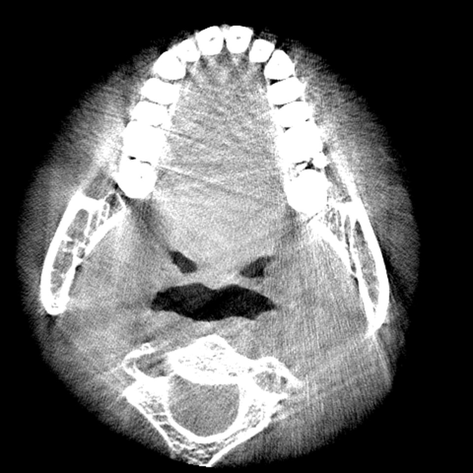
MDCT voxels for a head are generally larger in plane, and the slice thickness is often greater than the pixel size, so the voxels can be anisotropic, taller than they are wide, and coronal and sagittal reformats look blockier along the slice direction. What MDCT does better is soft tissue. Its fan beam collects far less scattered radiation than a flat panel catching a whole cone at once, so the contrast between muscle, fat, fluid and glands is real. On CBCT those tissues merge into a noisy mid grey (see CBCT artifacts).
Grey values versus Hounsfield units
A medical CT stores calibrated numbers. The Hounsfield scale sets water at 0 and air at about minus 1000, and a given tissue reads the same number on different days, in different parts of the field and, within reason, on different machines. That is what makes it possible to speak of bone or fluid as a number.
CBCT does not. The cone beam collects a lot of scatter, anatomy outside the reconstructed field still attenuates the beam, and beam hardening shifts values near dense objects. The result is a grey value that depends on where a structure sits, what is beside it and which machine took the scan. This is why JawAtlas shows a readout like "C 300 / W 1500 grey", never HU, and has no density readouts (see window, level and grey values). An imported medical CT is treated the same way: its intensities are shown as grey values, and judging density from them is not something the app does for either kind of scan.
Dose, in general terms
For a dental field of view a CBCT examination usually involves a lower dose than a medical CT of the same region. But the ranges overlap widely. Dose rises with the size of the field, the resolution and the exposure settings, and a large high resolution CBCT can approach a low dose CT protocol. The right comparison is between specific protocols on specific machines, and those figures belong in a machine's dose report, not a general guide.
Side by side
| Aspect | Cone beam CT | Multidetector CT |
|---|---|---|
| Beam and detector | Cone beam, flat panel, one rotation | Fan beam, detector rows, helical path |
| Patient position | Seated or standing | Lying on a moving table |
| Field of view | Cylinder sized to the region | Whole head routinely |
| Voxels | Small, usually isotropic | Larger, often anisotropic |
| Bone detail | Fine | Good, coarser |
| Soft tissue contrast | Poor | Good |
| Intensity scale | Uncalibrated grey values | Calibrated Hounsfield units |
| File format | DICOM | DICOM |
When each is used in teaching
CBCT is the natural teaching material for dental anatomy: roots, the mandibular canal, the sinus floor, impacted teeth and alveolar bone all read best on it, and the planes and conventions transfer directly to clinical viewers. Almost every guide on this site is written around a CBCT scan (see how to read a CBCT scan).
MDCT earns its place when the lesson is about soft tissue, the skull base or the whole head; when it is about Hounsfield units themselves, because you cannot teach a calibrated scale on an uncalibrated scan; and as a comparison, because a CT and a CBCT of the same region side by side is the fastest way to see what scatter and calibration mean. One of the openly licensed collections listed on this site is medical CT for that purpose (see free CBCT datasets).
How both open in JawAtlas
Both kinds of scanner write DICOM, and a series from either is a folder of slice files with the same technical tags for position, orientation and spacing (see what is DICOM). JawAtlas reads only a whitelist of technical tags, so patient identifiers never reach the app. The importer sorts the slices along the slice normal, checks for gaps and duplicates, and needs at least four slices. Supported transfer syntaxes are Implicit and Explicit VR Little Endian, Deflated, RLE Lossless and JPEG Lossless; lossy JPEG, JPEG 2000 and JPEG-LS files are rejected per file with a message. Import a folder through the Files app or a ZIP archive by AirDrop (see how to import DICOM from Files).
Once loaded, a CT and a CBCT behave the same way. The MPR panes use the DICOM spacing for the millimetre readouts, the Measure and Angle tools and the scale ruler, so an anisotropic CT measures correctly even though its reformats look coarser, and the edge labels come from the orientation cosines, so a tilted acquisition is still labelled correctly. Try the Soft Tissue preset first on a medical CT, because for once there is soft tissue to see.
Questions students ask
What is the difference between a CBCT and a CT scan?
Both build a three dimensional volume from X-ray projections. A cone beam CT uses a cone shaped beam and a flat panel detector in one rotation around a seated or standing patient. A medical CT uses a fan shaped beam and rows of detectors while the patient moves through the gantry on a table. CBCT gives finer bone detail in a smaller field; CT gives calibrated numbers and better soft tissue contrast.
Are CBCT grey values the same as Hounsfield units?
No. Medical CT numbers are calibrated so that water reads 0 and air about minus 1000, and they are comparable between scans. CBCT grey values are not calibrated and drift with scatter, position in the field and beam hardening. JawAtlas shows every scan's intensities as grey values for that reason.
Is a CBCT scan lower dose than a CT scan?
Usually, for a dental field of view, but the ranges overlap a great deal. Dose depends on the field of view, the resolution setting, the exposure settings and the machine, so a large high resolution CBCT can approach a low dose CT of the same region. Specific figures belong to the dose reports for a given machine, not to a general guide.
Can I open a medical CT scan in JawAtlas?
Yes, as long as it is a DICOM series in a supported transfer syntax with at least four slices. The importer sorts the slices along the slice normal and builds the volume from the DICOM spacing, so CT and CBCT behave the same way in the viewer. Intensities are shown as grey values in both cases.
JawAtlas is an education and training aid. It is not a medical device and must not be used for diagnosis, treatment planning or any clinical decision.
Last reviewed 2026-10-14